Skip to content

Sponsored Content Slideshow: Key considerations with digitial oral scanning

No quod sanctus instructior ius, et intellegam interesset duo. Vix cu nibh gubergren dissentias. His velit veniam habemus ne. No doctus neglegentur vituperatoribus est, qui ad ipsum oratio. Ei duo dicant facilisi, qui at harum democritum consetetur.
icon_howto
How-To
1 / 9

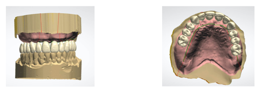
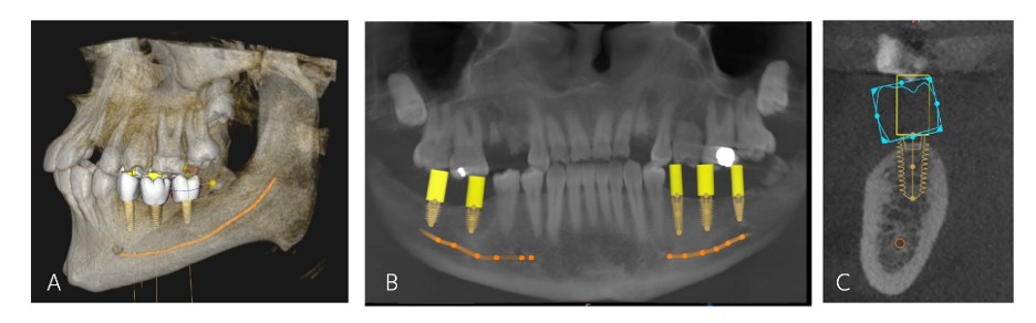
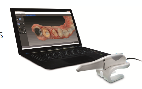
Dental impressions remain a critical step in restorative dentistry. Some of the advantages to optical impressions include the eliminations of pouring/storing plaster models, reduces patient discomfort, saves time during the impression process, simplifies the impression procedure1. In this sponsored Carestream slideshow, David A. Little, DDS, a practitioner, adjunct professor and consultant based in San Antonio, Texas guides you through implant placement using digital intraoral scanning.

1Mangano F. Gandolfi A, Luongo G, Ogozzo S. Intraoral scanner in dentistry: A review of the current literature. BMC Oral Health. 2017; 17(1):149.

2 / 9
This figure depicts a virtual wax-up for an edentulous implant-supported prosthesis. Understanding traditional approaches restorative dentistry is the first best step to transitioning to new technologies.
3 / 9
slidepic3
These photos let you compare a 2-dimensional versus 3-dimensional view of the same patient. The 2-dimensional image shows up to 30% distortion, while a cone-beam computed tomographic scan has zero distortion of hard tissue.
4 / 9
slidepic4
These three images offer a virtual plan for implant placement using cone-beam computed tomography. Photo B is of an intraoral standard triangle language file, and Photo C is an image of the planning software.
5 / 9
slidepic5
This series of slides offer a view of the virtual planning for a single tooth implant. Photo B: virtual wax-up. Photo C: Measurements for the location of anatomic structure. D: The surgical guide was fabricated from the digital intraoral scan obtained from the patient.
6 / 9
slidepic6
This radiograph is shown with reverse contrast. B: A photo of the scan body in place intraorally. C: The digital intraoral scan with the scan body in place is sent (electronically) to the dental laboratory.
7 / 9
slidepic7
This two photos show the intraoral scans with the healing abutment in place (A), and with the implant scan body in place to provide information for the position of the implant (B).
8 / 9
slidepic8

After the tooth is prepared and digitally scanned, the opposing dentition and a bite registration are obtained with a digital intraoral scanner. The dental laboratory will then use the digital impression (E) to print the model (F) and mill the crown.

9 / 9
slidepic9

For more information, download the free Carestream-sponsored ebook Integrating Digital Intraoral Scanning into General Practice.


Advertisement
LTGD


Related Posts