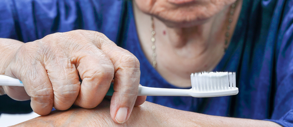
Facial fractures in older Americans
Chronic Falls are a frequent cause of injury among older Americans and contribute substantially to health care costs for this cohort. The incidence of facial fractures is on the rise in older cohorts, and hospitalization for such injuries may serve as an indication of a shift from a healthy to a compromised medical status. The aims of a retrospective epidemiologic study, published in the April issue of Dental Traumatology, were to provide data from US hospital emergency department (ED) visits for facial fractures and to investigate the outcomes and costs associated with these visits.
Data were obtained from the National Emergency Department Sample (NEDS), a 20% stratified sample of all US hospital ED visits from 2008 through 2014 for visits with an International Classification of Diseases, Ninth Revision, diagnosis code of facial fracture. The inclusion criterion was 65 years and older. Variables examined included age, sex, annual household income quartile, insurance type, hospital region, hospital ownership, and teaching status. Both ED and hospital stay charges were adjusted for inflation to 2014 US dollars. The primary outcome variable was mortality after the ED visit, evaluated by mortality during the ED visit and during hospitalization after the ED visit. The year of the study was the primary independent variable. Simple descriptive statistics summarized the data, and a trend analysis was carried out to determine whether hospital-related mortality was altered during the study. Statistical analysis was performed using SAS 9.4 Version software (SAS Institute) and 2-sided test, and a P value of .05 set as statistically significant.
A total of 540,748 US hospital–based ED visits for facial fractures among patients 65 years and older was recorded during the study. The greatest number of fractures occurred in the cohort aged 75 through 84 years (38.4%), followed by those aged 65 through 74 years (32.7%) and those 85 years and older (28.9%). Facial fractures were most common in women (62.7%). Medicare was the most frequent payer (85.2%), followed by private insurance payers (10.5%); only 1.4% of the ED visits were incurred by patients with no insurance. Hospitals in the South treated 38.4% of patients seen in EDs for facial fractures. Overall, large metropolitan areas accounted for nearly one-half (47.9%) of ED visits. Teaching hospitals treated 43.1% of the patients, while only 15% visited EDs at level 1 trauma center–designated hospitals.
The most frequent type of fractures were closed fractures of nasal bones (61.3%), closed fractures of other facial bones (16.7%), closed blowout fractures of the orbital floor (15%), closed fractures of the malar and maxillary bones (14%), and open fractures of the nasal bones (3.5%). Falls were the predominant cause of fracture (81.8%), followed by motor vehicle accidents (4.5%) and being struck by others (3.9%). A total of 64.1% of patients were seen in the ED and released, while 35.6% were admitted to hospital. The average charge per ED visit was $5,507, and the mean hospitalization charges for inpatients was $61,321 with an average stay of 5.5 days. With regard to mortality, 633 patients (0.1%) died in the ED, while 1.5% died after hospital admission. There were no statistically significant trends in ED mortality rates or death after admission rates during the study.
In this study, falls were the most prevalent cause of facial fracture among older adults in the United States at nearly 82%, with a cost of $12.5 billion, of which 85% was paid for using public (Medicare) dollars. The authors advocate implementation of multifactorial fall prevention strategies, including at least 3 of the following: education, risk assessment and suggestion, exercise, medical care, hazard assessment, and modification. The researchers were encouraged that mortality rates held constant during the study and speculate this may have been due to the institution of multifactorial interventions in some locations. One limitation of the study was the lack of opportunity to perform chart abstraction to examine variables such as causes for hospital admissions and duration of stay, circumstances of falls, ratio of nursing home residents, and association of facial fractures with elder abuse. The authors suggest future studies should examine these variables and include a more detailed analysis of the different types of facial fracture and their outcomes. On a practical note, dental teams should be mindful of potential trip hazards (rheostats, sensor cords) in operatories and place them out of the patient footpath areas to reduce the incidence of falls during dental visits.
Read the original article here or contact the ADA Library & Archives for assistance.
Hallmarks of frailty in older adults
In Frailty is a term used to describe older adults considered weaker and more vulnerable than their age-matched cohorts, despite similar conditions, demographics, and sex. Several indexes for the clinical diagnosis of frailty exist, yet their ability to identify progression on the spectrum of robust to prefrail to frail is limited. The aim of this review published online February 19 in Clinical Interventions in Aging was to summarize the evidence on the utility of currently used biomarkers for frailty and compare these with upcoming biomarkers with respect to their strengths, weaknesses, validity, and predictive value.
Currently used circulating biomarkers for frailty include inflammatory mediators, markers of clinical parameters, hormones, products of oxidative degradation, and levels of antioxidants. Unfortunately, these biomarkers are also biomarkers of the aging process itself, only reflect single aspects of the frailty complex, and are poor predictors of condition progression. Increasing numbers of frail people as a result of global aging lend import to the need to find useful biomarkers so early diagnosis and intervention can improve clinical outcomes.
Inflammation is associated with the aging process, and levels of several inflammatory markers, notably interleukin-6, tumor necrosis factor , and C-reactive protein have been shown to correlate with several components of frailty syndrome and are simple to measure. However, their lack of utility in identifying the phase of a patient on the frailty spectrum, inability to predict progression, and general increase in level during the normal aging process render them unsatisfactory as biomarkers for frailty.
As many of the clinical manifestations of frailty mirror the body’s musculoskeletal profile, hormones such as testosterone and its precursor, dehydroepiandrosterone, parathyroid hormone, Vitamin D and insulinlike growth factor 1 have been investigated as possible biomarkers for frailty. Hormonal biomarkers are in widespread use, can assist in diagnosis, and may predict progression along the frailty spectrum. However, they can be challenging biomarkers with variability in levels across seasons and ethnic groups. Further research would be necessary to establish normal reference values for all populations and identify their utility in predicting onset in these patients with complexities and the management of their treatment.
Metabolic dysregulation, with progression to diabetes, has emerged as an increasing public health concern globally. Glycated hemoglobin estimates how much glucose a red blood cell has encountered during its 3-month life span and has been shown to be indicative of frailty. However, it has not yet been shown to predict progression along the frailty spectrum and has not been correlated with poorer clinical outcomes. Several studies have found a correlation between a decrease in circulating hemoglobin, albumin, and low glomerular filtration rate and frailty. However, these biomarkers are also present in comorbidities frequently seen in older adults, rendering their use as stand-alone markers of frailty suspect. Similarly, increased levels of oxidative markers and decreased levels of antioxidants are associated with common comorbidities and with the normal aging process itself. Thus, the flaws in and confounding nature of the currently available biomarkers drives research to finding better tools to diagnose and predict progression of frailty.
Circulating mesenchymal stem cells can aid in repair of damaged tissues, but, depending on their tissue source, demonstrate great variability in differentiation capacity and gene-expression profiles. Circulating osteoprogenitor (COP) cells, which act as surrogates for the marrow stem cell population, freely flow across the endothelial barrier and appear to have the potential to serve as biomarkers for musculoskeletal disorders. In 1 study, lower COP percentage showed strong correlation with clinical indicators of frailty. Lamin A is an intermediate filament present in the nuclear lamina, and patients with deficient lamin A develop musculoskeletal disease. Low lamin A levels in COP cells have been strongly associated with frailty. Longitudinal studies are required to evaluate the sensitivity, specificity, and predictive value of COP percentage and lamin A-COP as biomarkers for frailty. Paralleling the global increase in life expectancy will be a concomitant rise in the incidence of frailty, prompting the need for biomarkers to enable early diagnosis and intervention, allowing older adults to remain healthy and independent longer.
Read the original article here or contact the ADA Library & Archives for assistance.
Advertisement
Delivery strategies for oral health care to patients with dementia in nursing home settings
Care-resistant behaviors (CRBs) are reported by nursing home (NH) staff as a significant deterrent to the provision of oral hygiene to residents with dementia. The aim of this randomized, repeated measures clinical trial was to examine the utility of Managing Oral Hygiene Using Threat Reduction (MOUTh), a nonpharmacologic, relationship-based intervention, versus usual methods of oral care delivery (control) in reducing the occurrence and intensity of CRBs and improvement of oral health in NH residents with dementia who resisted oral hygiene. Secondary outcomes included the duration of the oral hygiene event and whether oral hygiene activities were completed during the event. The results of the study were published in the December 2018 issue of Gerodontology.
Patients were recruited from 9 U.S. NHs, and consent was obtained from legally authorized representatives. Inclusion criteria included age 55 years or older, dentate with at least 2 adjacent teeth or edentulous and using a complete denture in at least 1 arch, diagnosis of dementia, and identified by staff as being resistant to oral care. Forty-six patients in the experimental group and 54 in the control group contributed data to the project analysis. Trained research assistants blinded to treatment assignments were assigned to provide oral hygiene.
Oral care for dentate patients consisted of brushing all teeth and the tongue with a soft toothbrush and fluoride toothpaste followed by use of interdental brushes and an antimicrobial mouthrinse. Oral care for edentulous patients included use of a soft toothbrush, denture cleaning paste and a denture brush.
All study participants were observed receiving oral care from NH staff twice per day for 7 days to establish baseline levels of CRBs. On day 8, participants who were randomly assigned to experimental or control groups received oral care twice per day for 3 weeks from research assistants. In the experimental group, the MOUTh strategies used included establishing rapport with patient by approaching him or her at or below eye level with a calm, pleasant demeanor, avoiding elder speak, chaining, cueing, distraction, bridging, rescue, and hand-over-hand method.
Occurrences of CRBs were assigned a binary score (behavior present or not). CRB intensity was assessed with the revised Resistiveness to Care Scale. Participants’ oral health was assessed using the Oral Health Assessment Tool (OHAT). Duration of oral hygiene was measured with a stopwatch. Completion of oral care was assigned a binary score (completed or not).
During the 3-week intervention period, participants in the experimental group had twice the odds of assenting to oral care and completing oral care, both of which were relevant and significant, as did participants in the control group. The duration of oral care increased for both groups in the intervention period, but it was higher by 0.72 minutes in the experimental group, a statistically significant result. During the intervention period, the intensity of CRBs decreased in both groups. The decrease was greater in the experimental group, a result which was considered positive, although it was not statistically significant. Likewise, OHAT scores were better for both groups as a result of intervention, slightly more so for the experimental group, but again, not statistically significant.
The authors point out that OHAT scores showed clinical improvements for both groups during the intervention period, which may indicate that oral hygiene is not considered a priority in NH settings. They note that this is the first randomized controlled trial of a nondrug–based intervention designed to decrease CRBs and improve oral health measures in patients with CRBs and dementia, a group that has typically been excluded in prior studies. The research assistants providing experimental group oral care were trained to implement strategies to prevent escalation of CRB severity. Thus management, rather than prevention of CRBs, appears to be responsible for improved oral care in patients with dementia. The authors suggest future studies evaluate optimal frequency of oral care activities, as well as protocols, for patients who have dysphagia and testing the efficacy of the MOUTh protocol in patients with dementia who reside in assisted living homes.
Read the original article here or contact the ADA Library & Archives for assistance.
Therapeutic approaches to salivary hypofunction
The subjective sensation of dry mouth is termed xerostomia, and hyposalivation is measured objectively as a reduction in salivary flow, accompanied by changes in the composition of saliva. Researchers conducted a MEDLINE/PubMed search to examine current therapeutic strategies for salivary hypofunction. The results of their study were published in the December 2018 issue of Gerodontology.
For the review, the authors conducted a search of the English-language literature from 1995 through 2017. Treatments for xerostomia were categorized as symptomatic, topical or systemic stimulants, and regenerative. Twenty-five clinical trials were included, of which 24 were randomized controlled trials.
Increased fluid intake and use of salivary substitutes proved to be the most common symptomatic approaches to treating xerostomia. Salivary substitutes affect the oral mucosa directly, but do not enhance production of saliva. Three controlled clinical trials were selected to investigate the effects of salivary substitutes on xerostomia. No evidence was found that hydroxymethyl cellulose, polyglycerylmethacrylate, or xanthan gum relieved symptoms of xerostomia.
Citric acid and malic acid acidify the mouth, prompting salivary stimulation. In the 3 randomized controlled clinical trials, both citric and malic acid improved xerostomia, but the sample sizes of the trials were small and the duration of follow-up was short. In addition, use of the acids may make patients prone to dental hypersensitivity, erosion, and caries. In 2 randomized clinical trials, no evidence was found to support a positive effect of sugarless chewing gum on hyposalivation. Three studies suggested that acupuncture was efficacious in relieving xerostomia. Four of 5 studies indicated a beneficial effect of pilocarpine on symptoms of hyposalivation in patients with radiation-induced xerostomia and Sjögren syndrome. However, as a result of being a cholinergic drug, pilocarpine can produce negative side effects. Two randomized controlled clinical trials did not support effectiveness of bethanechol in reducing xerostomia. Four trials demonstrated that cevimeline was useful in symptomatic and functional improvement for patients with Sjögren syndrome and those who had undergone radiation but caused nausea.
In 3 studies conducted in irradiated rats, transplanted stem cells from bone marrow or adipose tissue significantly ameliorated salivary gland dysfunction. Animal studies also demonstrated promise of gene therapy in modulating radiation-induced salivary dysfunction. Sufficient evidence was not found for the use of hyperbaric oxygen in the treatment of radiation-induced xerostomia.
Xerostomia causes significant suffering globally, and approaches to the management of salivary hypofunction are wide-ranging. Thorough review of the literature led the authors to observe that pilocarpine and cevimeline have the most pronounced efficacy in treating salivary hypofunction, but often produce negative gastrointestinal side effects. The use of citric and malic acids increase salivary flow but can cause dental erosion and caries. The use of salivary substitutes, chewing gum, bethanechol, and hyperbaric oxygen do not appear to be effective in ameliorating salivary hypofunction. The authors propose that randomized controlled clinical trials in humans be conducted to evaluate the utility of acupuncture, stem cell therapy, and gene therapy in relieving xerostomia.
Read the original article here or contact the ADA Library & Archives for assistance.

Advantage Arrest SDF 38%
Advantage Arrest is the first and only American-made silver diamine fluoride (SDF), a must-have for every dental office. Advantage Arrest is the only tinted SDF formula, enhancing placement visualization, and is available in an economical 8 mL bottle and unit-dose packaging for enhanced asepsis. Advantage Arrest is an immediate and effective desensitizer (D9910), a discloser of incipient lesions (D1354), and a proven caries preventive agent (D1208). Elevate Oral Care preventive care consultants have conducted educational staff meetings for thousands of oral health professionals since launching Advantage Arrest in 2015. To purchase Advantage Arrest directly and/or schedule your informative staff meeting today click here.
Live Patient Botox and Dermal Fillers courses at ADA Headquarters  There are still seats available for Botulinum Toxins and Dermal Fillers for Every Dental Practice. Join us May 10-11, 2019 at ADA Headquarters in Chicago to learn Botox and Dermal Filler treatment techniques that you will be able to immediately implement into your practice, led by Dr. Louis Malcmacher and his team at the American Academy of Facial Esthetics. Register now!

The consulting editor for JADA+ Scan — Healthy Aging is Linda C. Niessen, DMD, MPH; Dean and Professor; Nova Southeastern University College of Dental Medicine. |
|


