
Increasing root canal taper predisposes endodontically treated maxillary molars to fracture, but access cavity design does not play a significant role, according to a study in the September issue of Journal of Endodontics.
“Tooth fracture is 1 of the most undesirable phenomena in endodontically treated teeth (ETT) and usually leads to tooth extraction,” researchers wrote.
They also wrote, “The endodontic access cavity is considered an important step in endodontic treatments.” However, they pointed to the loss of tooth structures due to therapeutic endodontic procedures—such as access cavity and root canal preparation—as predisposing factors associated with tooth fractures.
In their investigation, the researchers aimed to quantify the effect of access cavity design and root canal taper on fracture resistance in ETT. They focused on maxillary molars because available data on fracture resistance of maxillary molars was scant, unlike that for mandibular molars.
The study discussed the differences between traditional access cavity (TAC) and the newer conservative access cavity (CAC), citing TAC as drawing some criticism owing to the possibility of excessive tooth removal and describing CAC, inspired by concepts of minimally invasive dentistry, as slow to impact the endodontist mainstream practice and not well supported by research data.
Researchers worked from 2 null hypotheses in evaluating and comparing the effect of access cavity preparation and 3 different root taper preparations on ETT fracture resistance of maxillary molars. The first null hypothesis was there would be no difference in fracture resistance of teeth with different access cavity designs. The second null hypothesis was there would be no difference in the fracture resistance of roots with different root canal tapers.
Collectively, researchers evaluated a total of 78 sound maxillary first and second molars extracted for periodontal reasons. They divided the molars into 2 experimental groups: 1 for tapering assessment and 1 for cavity preparation approach assessment.
In the tapering assessment group, the researchers randomly assigned 30 sound distobuccal maxillary molar roots to 1 of 3 groups (n = 10): a .04 taper, a .06 taper, or a .08 taper. After preparing the specimens for endodontic treatment, the investigators used a rotary system for instrumentation of canals.
In the cavity preparation group, the researchers randomly assigned 48 intact maxillary first and second molars to 1 of 3 groups (n = 16): intact teeth, TAC, and CAC. In this group, they tested fracture resistance using a universal testing machine.
The investigators used statistical software to analyze results. They applied statistical test of normality—the Shapiro-Wilk test. They set level of importance for all comparisons as P ≤ .05 and used the Tukey test as the post hoc evaluation.
In the root canal tapering group, the .04 taper had the highest fracture resistance (average [standard deviation {SD}], 259.61 [52.06]), while the .08 taper head the lowest fracture resistance (average [SD], 168.43 [59.63]). The .04 and .06 taper groups did not significantly differ from each other (P > .05), although both groups differed significantly from the .08-taper group (P ≤ .05).
From their evaluations, researchers concluded that increased root canal tapering and access cavity preparation decrease fracture resistance. The newer access cavity design—CAC—showed no significant impact on improving fracture resistance compared with TAC.
Read the original article here or contact the ADA Library & Archives for assistance.
Advertisement

Infection control and apical periodontitis
A triple antibiotic solution significantly improved root canal disinfection and was at least as effective in controlling infection in teeth with apical periodontitis as a calcium hydroxide/chlorhexidine paste, according to researchers in a study published in the October issue of Journal of Endodontics.
“Because bacterial infection is the primary cause of the different forms of apical periodontitis, the successful outcome of endodontic treatment procedures relies on effective infection control,” researchers wrote. “However, adequate disinfection of the root canal system of teeth with pulp necrosis and apical periodontitis remains a crucial challenge to be overcome in endodontics.”
Researchers cited a need for finding better, more predictable medications for disinfecting the root canal system as studies have shown that bacteria can still be present in canals even 1 week after endodontic treatment despite calcium hydroxide medication.
In the randomized clinical study, researchers sought to compare the clinical antibacterial effectiveness of endodontic treatment protocols using either a triple action solution or calcium hydroxide/chlorhexidine paste as interappointment medication in infected canals of teeth with primary apical periodontitis. The researchers evaluated the reduction of total bacterial levels using the culture-independent molecular microbiology assay quantitative real-time polymerase chain reaction.
To conduct their study, researchers included 48 participants in their study population, including 23 females and 25 males ranging in age from 13 to 71 years (mean, 41 years). They were patients who had received treatment of apical periodontitis associated with mature teeth from January 2016 through October 2017 at the endodontic clinic at the School of Dentistry, Estacio se Sa University, Rio de Janeiro, Brazil. Inclusion criteria called for teeth confirmed to have a single root and a single canal presenting with a carious lesion and necrotic pulp. Exclusion criteria were teeth with extensive crown destruction by caries that could not be restored or isolated with a rubber dam, root or crown fracture, previous endodontic treatment, symptoms, periodontal pockets measuring more than 4 millimeters deep, and patients who received systemic antibiotic therapy over the previous 3 months. Also, participants had no significant systemic conditions.
The primary outcome evaluated was a reduction in intracanal bacterial count after using treatment protocols with either an antibiotic mixture or a calcium hydroxide paste as interappointment medication. Researchers used software to calculate the sample size and arrived at a minimum of 21 teeth per group to sufficiently show a 5% difference in bacterial count with a power of 90%. Teeth were randomly assigned to 2 groups of 24 each according to the medication used. Randomization was achieved by drawing slips of paper with the group name from a bowl after undergoing chemomechanical procedures and immediately before application of the medication.
After collecting samples from the root canals under strict asepsis, researchers took an initial sample (S1) before chemomechanical procedures from the root canals to serve as the baseline. Another sample was taken after chemomechanical preparation (S2), and a third after intracanal medication (S3). Researchers evaluated DNA extracts from clinical samples for total bacterial reduction using a 16S ribosomal RNA gene-based quantitative polymerase chain reaction assay.
Bacteria were ultimately found in all S1 samples, with significant reduction posttreatment (P < .01). There was not a significant difference between bacteria levels in S2 and S3 samples (P > .05). Bacterial reduction for S2 through S3 was 97% in the antibiotic group and 39% in the calcium hydroxide/chlorhexidine group.
Researchers determined that triple antibiotic solution concentrated at 1 milligram per milliliter had strong antibacterial effects in vivo and should be considered an alternative intracanal medicament.
Read the original article here or contact the ADA Library & Archives for assistance.
Advertisement


CBCT, apical periodontitis and root-filled teeth versus nonroot-filled teeth
The accuracy of cone-beam computed tomography (CBCT) in diagnosing apical periodontitis (AP) depends on the status of the tooth, with higher accuracy possible in nonroot-filled teeth than root-filled teeth, researchers found in a study on the topic. They published their results online September 28 in International Endodontic Journal.
“Several studies have been performed to assess the diagnostic accuracy of CBCT for detection of periapical lesions, and the overall conclusion was that more periapical radiographic [PR] lesions were identified using CBCT compared with PR,” researchers wrote.
However, they found that most of the studies did not use a valid reference standard or were in vitro studies evaluating induced (artificial) bone lesions, disqualifying results for direct clinical application.
For their study, the researchers aimed to assess the accuracy of CBCT in diagnosing AP using histopathology as a reference standard in ex vivo human jaws with a comprehensive and diverse sample of teeth. Researchers declared it as important that the study design included teeth with a variety of dental treatments and diseases, including both root-filled and nonroot-filled teeth. “It has been shown that several parameters increase the risk of AP in a tooth, in particular root filled teeth have been shown to have AP more often than non-root filled,” they wrote.
They obtained individual dentate half maxilla or half mandible specimens that included soft tissues and conducted radiographic examinations—both PR and CBCT—of all dentate areas of the jaw specimens.
Researchers identified 223 teeth with 340 roots based on PR for the study. For each tooth, they recorded the identification number, jaw, tooth number, number of the radiographically visible roots, root canal treatment, intrapulpal post, coronal restoration (restoration, crown/bridge abutment), caries, marginal bone level, and periapical status. The teeth reflected a sample from all tooth groups and with different diseases and treatment statuses. Researchers processed the study teeth for histopathologic analysis.
Researchers performed CBCT, and 3 observers assessed the presence of AP using the probability index. As a reference standard to estimate diagnostic accuracy, researchers conducted histopathologic examination of the periapical area.
Significantly, nonroot-filled teeth had overall high estimates of diagnostic accuracy across sensitivity, specificity, positive predictive value, and negative predictive value, whereas all estimates were lower for root-filled teeth.
Researchers concluded that, depending on the treatment status of the tooth, the diagnostic accuracy of CBCT is high and almost all cases of AP can be diagnosed correctly with just a small risk of overdiagnosis in nonroot-filled teeth. Conversely, in root-filled teeth all diagnostic accuracy parameters were lower leading to less accuracy of AP diagnosis on these teeth using CBCT.
Read the original article here or contact the ADA Library & Archives for assistance.
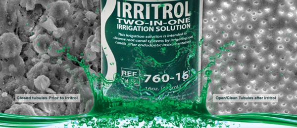
Irrigate and disinfect with Irritrol
Endodontic disinfection can be challenging. Irritrol eliminates the need for separate step irrigation of EDTA and CHX irrigation. Irritrol is proven to irrigate and rapidly disinfect the root canal. Independent testing showed that Irritrol had a disinfection rate of 99.99%* (Independently confirmed by Nelson Labs; Time kill study protocol #STP01). See why Irritrol won the award from Dental Advisor for Top Endodontic Irrigant. Read more.

Registration for AAE19 is now live ![]() The American Association of Endodontists is proud to bring its annual meeting to the beautiful city of Montréal, April 10-13, 2019. Register by March 1 to save! Learn more at www.aae.org/aae19.
The American Association of Endodontists is proud to bring its annual meeting to the beautiful city of Montréal, April 10-13, 2019. Register by March 1 to save! Learn more at www.aae.org/aae19.
Over 50 hours of captured content at ADA 2018  Relive your ADA meeting experience, or take a course that you missed! Didn’t attend the Annual Meeting? Now’s your chance to experience the top ADA 2018 speakers from the comfort of your own home. Many courses from ADA 2018 will become available soon, subscribe now to start your one-year, all-access pass to ADA CE Online. Your subscription includes everything in the ADA CE Online library as well as all JADA and new courses being added. For more information, visit ada.org/cesubscriptions.

The consulting editor for JADA+ Specialty Scan — Endodontics is Dr. Susan Wood, Diplomate, American Board of Endodontics. |
|
