
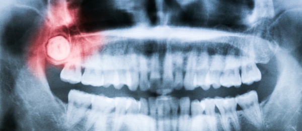
Radiographic predictors of bone exposure in patients with medication-related osteonecrosis of the jaw
Medication-related osteonecrosis of the jaw (MRONJ) is an adverse effect of antiresorptive and antiangiogenic treatment for patients with osteoporosis or bone malignancies. The aim of this retrospective study was to assess the cone-beam computed tomographic (CBCT) scans of 23 patients initially diagnosed with stage 0 MRONJ and correlate their radiographic parameters with subsequent bone exposure (BE). The study was published online August 29 in Oral Surgery Oral Medicine Oral Pathology Oral Radiology.
From January 2013 through April 2015, 23 patients (18 female, 5 male) with a history of treatment with bisphosphonates, denosumab, or both were referred by their general dentists to the Oral and Maxillofacial Surgery Clinic in the School of Dentistry at the University of California, Los Angeles. None of the patients had BE or fistula formation at the initial visit. Their nonspecific signs and symptoms included dull bone pain, altered neurosensory function, mucosal erythema, and edema.
All patients underwent panoramic radiography and CBCT, and intraoral photographs also were obtained. The mean follow-up period was 10 months (range, 1 month-3 years). Radiography was performed using a 3D Accuitomo 170 scanner (J Morita USA) with the following parameters: 90 kilovolt (peak) and 6 milliamperes with a 17.5-second exposure during 360-degree rotation. The field of view was 6 × 6 centimeters with a 0.125-millimeter isometric voxel or 10 × 14 cm with a 0.25-mm isometric voxel.
One oral and maxillofacial surgeon performed all clinical examinations. A senior resident in oral and maxillofacial radiology and a board-certified oral and maxillofacial radiologist evaluated all CBCT scans.
The investigators evaluated 5 radiographic parameters at the initial visit: trabecular sclerosis, cortical erosion, periosteal reaction, sequestration, and craterlike defects. They classified the findings as absent (0), localized (involving the area of 1 tooth: value = 1), or extensive (exceeding the area of 1 tooth: value = 2). They also calculated a composite radiographic index (CRI), which is the sum of the values for the 5 parameters.
Radiographic evaluation revealed extensive trabecular sclerosis in 13 of the 23 patients (57%) and localized trabecular sclerosis in 7 patients (30%). The investigators observed extensive cortical erosion in 3 patients (13%) and localized erosion in 13 patients (57%). The radiographic findings also revealed extensive periosteal reaction in 2 patients (9%) and localized periosteal reaction in 3 patients (13%). Of the 23 scans, 8 (35%) exhibited extensive sequestrum formation and 3 (13%) exhibited localized sequestrum formation. The researchers observed an extensive craterlike defect on 3 scans (13%) and a localized craterlike defect on 13 scans (57%).
All patients were treated conservatively. On clinical follow-up, the oral and maxillofacial surgeon examined patients for progression to clinical BE (MRONJ stages 1-3). Of the 23 patients, 3 experienced resolution of symptoms and abnormalities and were classified as having dental disease. Ten patients had persistent pain and abnormal clinical findings on follow-up, but no evidence of BE. They were classified as having stage 0 MRONJ with no BE (NBE). The remaining 10 patients had clinical BE (CBE) and inflammation or infection of the surrounding soft tissues. Three of these patients progressed to stage 1 MRONJ, 5 progressed to stage 2, and 2 progressed to stage 3 (extraoral fistula formation).
Patients in the dental disease group exhibited minimal radiographic findings, and their CRI was significantly lower than that of patients in the NBE (P < .05) and BE groups (P < .001). Patients in the BE group had a significantly higher CRI than those in the NBE group (P < .05). Moreover, the initial CBCT scans of 9 of 10 patients in the BE group exhibited signs of sequestration, whereas the initial scans of 8 of 10 patients in the NBE group did not exhibit signs of sequestration.
The study findings show that radiographic evidence of sequestrum formation was an important predictor of progression from stage 0 MRONJ to clinical BE. The authors concluded that all patients who have received antiresorptive or antiangiogenic medications and exhibit abnormal clinical symptoms or signs should undergo a CBCT examination.
Read the original article here or contact the ADA Library & Archives for assistance.
Advertisement
Assessing risk factors for external root resorption in second molars associated with impacted third molars
External root resorption (ERR) or caries can be observed on the distal surface of a second molar adjacent to an impacted third molar. The aim of this retrospective cross-sectional study was to examine the risk factors for ERR in second molars associated with impacted third molars. The study was published online September 3 in Journal of Oral and Maxillofacial Surgery.
The study sample was composed of 200 patients (118 women, 82 men) seen in the Department of Oral and Maxillofacial Radiology at Necmettin Erbakan University in Konya, Turkey, from 2013 through 2017. Their mean (standard deviation) age was 27.26 (8.60) years. All patients underwent cone-beam computed tomography (CBCT) for diagnostic purposes (such as third-molar extraction, orthodontic evaluation) and had at least 1 impacted third molar. The researchers randomly selected 1 impacted third molar in each patient for study inclusion.
Inclusion criteria were age 18 years and older, eruption of all teeth except third molars, and at least 1 impacted (or partially erupted) third molar. Exclusion criteria were completely erupted third molars with cystic or tumor lesions associated with impacted teeth; third molars with evidence of less than two-thirds root development; second molars with severe carious lesions, crowns, or distal restorations; or any artifacts.
The researchers used a 3D Accuitomo 170 unit (Morita) to obtain the CBCT images. One radiologist evaluated all sagittal and axial CBCT slices for the presence of ERR on the second molar. The researchers diagnosed ERR according to Al-Khateeb and Bataineh’s definition: a “clear loss of substance in the root of adjacent mandibular second molar due to direct contact between it and the impacted third molar.” They also categorized ERR according to its location (cervical, middle root third, or apical root third) and severity (slight, moderate, or severe).
The investigators distinguished ERR from dental caries based on radiologic appearance. If there was a clear gap between the second molar and the crown of the third molar, and radiolucency showed irregular morphology, the diagnosis was caries.
Of the 200 impacted third molars, 42 (21%) were associated with ERR. Logistic regression analysis showed a significantly greater prevalence of ERR in mandibular second molars (n = 37) than in maxillary second molars (n = 5). The risk of experiencing ERR was about 4 times greater in mandibular molars than in maxillary molars (P < .05). Moreover, the study findings showed that mesioangular or horizontal inclination of third molars was associated (P < .05) with a greater prevalence of ERR compared with distoangular or vertical inclination.
The study findings revealed no statistically significant relationship between sex and the presence or severity of ERR (P > .05). Although age was not an independent risk factor for ERR, the study results showed that resorption severity increased with age (P < .05), the authors wrote.
One study limitation was the use of radiologic diagnosis exclusively rather than correlation of radiologic findings with clinical findings. The researchers suggested that direct visualization of ERR after tooth extraction along with histopathologic analysis could be helpful.
In light of the study results, decisions regarding extraction of impacted third molars should take into consideration their location (mandible or maxilla) and inclination (mesioangular or horizontal versus distoangular or vertical), the authors wrote. They concluded that future studies involving clinical and histologic evaluation are needed to confirm the results of this cross-sectional retrospective study.
Read the original article here or contact the ADA Library & Archives for assistance.
Examining the prevalence and characteristics of the infraorbital canal and Haller cells in edentulous patients
Successful implant treatment in the anterior maxilla requires familiarity with the infraorbital region and its possible variations. In this retrospective study, researchers evaluated the prevalence and characteristics of the infraorbital canal and Haller cells (infraorbital ethmoidal cells) in edentulous patients. The study was published online July 8 in BioMed Research International.
The study sample comprised 291 diagnostically acceptable panoramic radiographs of edentulous patients (103 men, 188 women) randomly selected from the archives of the Oral and Maxillofacial Radiology Department at Eskișehir Osmangazi University in Eskișehir, Turkey. The mean (standard deviation) age of patients was 66.63 (10.11) years, with a range of 38 through 88 years.
The radiographs were acquired with a digital unit (ProMax, Planmeca) set to the following parameters: 64 kilovolts, 6 milliamperes, and 16-second duration. Two oral and maxillofacial radiologists independently viewed the first 60 radiographs on the same computer under optimal lighting conditions. Interrater reliability was high (κ = 0.86 for Haller cells, κ = 0.89 for the infraorbital canal).
The investigators grouped the radiographic characteristics of the canal borders according to Scarfe and colleagues’ classification:
- Type I: Both the borders of the orbital and antrum parts of the canal are radiopaque.
- Type II: Both the borders of the orbital and antrum parts of the canal are invisible with no linear radiopacity.
- Type III: The orbital part of the canal is radiolucent with no linear radiopacity; the antrum part of the canal is radiopaque with linear radiopacity.
The investigators grouped the Haller cells according to the number and shape of loculations: multilocular, unilocular with septae (clustered minor locules), or unilocular (without septae). In addition, they identified Haller cells according to Ahmad and colleagues’ criteria:
- Well-defined, round, tear-drop–shaped radiolucency, single or multiple, unilocular or multilocular, with a smooth border, which may or may not be corticated.
- Located medial to the infraorbital foramen.
- All or most of the border in the panoramic section is visible.
- The inferior border of the orbit lacks cortication or remains indistinguishable in areas superimposed by this entity.
The study findings showed that the infraorbital canal was visible in 246 of the 291 radiographs (84.5%); 203 of these cases were bilateral and 43 were unilateral (equal for the right and left sides). On the right side, 116 radiographs (50%) exhibited Type III canals; on the left side, 94 radiographs (44%) exhibited Type III canals. The authors did not observe any significant differences between men and women regarding the visibility of infraorbital canals.
Of the 291 radiographs, 69 (23.7%) exhibited a total of 88 Haller cells, the authors wrote. Fifty of the 69 cases were unilateral, and 19 were bilateral. Among the 88 presentations, 36 on the right side were unilocular, 4 were multilocular, and 4 were unilocular with septae (clustered); on the left side, 37 were unilocular, 1 was multilocular, and 6 were unilocular with septae. Of the 19 bilateral cases, 14 (74%) were unilocular. As with the infraorbital canals, the study results did not show any significant differences between men and women with regard to the prevalence of Haller cells.
The researchers pointed out 2 advantages to using panoramic radiography to identify infraorbital canals and Haller cells: anatomic structures over a large area can be visualized with a single dose of low radiation, and the projection angle of panoramic radiography enables the viewer to observe the scope of the infraorbital canal. However, 1 disadvantage of panoramic radiography is its lower sensitivity compared with that of 3-dimensional imaging.
The researchers suggested that future studies focus on the prevalence of Haller cells in patients with unexplained orofacial pain. Moreover, oral and maxillofacial radiologists and surgeons need to use cone-beam computed tomography to perform in-depth studies of the course of the infraorbital canal in the maxillary sinus and its relationship with sinus septae and Haller cells.
Read the original article here or contact the ADA Library & Archives for assistance.
Radiographic evidence of dermal fillers injected decades earlier in 3 elderly patients
Dermal filler procedures have become increasing popular in the past several years. These fillers can appear as radiopaque shadows on radiographic images. In this case series, published in the September issue of Imaging Science in Dentistry, the authors described 3 elderly patients with radiographic evidence of dermal fillers that had been injected decades earlier.
Case 1. An 88-year-old edentulous woman was seen in the Department of Oral and Maxillofacial Surgery at Kyungpook National University Dental Hospital in Daegu, South Korea, for implant placement and removal of flabby tissue in the anterior maxillary region. A panoramic radiograph (Orthopantomograph OP100 D, Instrumentarium Imaging) revealed a cluster of discrete radiopacities in the left and right cheek regions. On questioning, the patient acknowledged having been treated twice with subdermal injections of filler in both cheeks and zygomatic areas about 40 years earlier.
The authors performed cone-beam computed tomography (CBCT) using a Pax-Flex 3D unit (Vatech) with a 120- × 85-millimeter field of view, 90 kilovolt (peak), and 10 milliamperes. The CBCT images revealed multiple calcifications in the soft tissue of the cheeks and retromaxillary spaces bilaterally. A few of the opacities were larger than 11 mm in diameter and were surrounded by a thin radiopaque rim. The calcified lesions were distinct from the adjacent bony structures, and the surrounding soft tissues appeared intact. The authors concluded that the calcifications were either a result of the long-standing presence of the cosmetic filler itself or a secondary reaction to the filler.
Case 2. A 91-year-old woman was seen for treatment of osteomyelitis of the anterior mandible. The physical examination revealed a purulent discharge with exposed necrotic bone in the anterior mandibular region. A panoramic radiograph showed ill-defined osteolytic bony changes with internal radiopacities and surrounding sclerosis. In addition, multiple small radiopacities were observed in both cheeks and on the right side of the chin.
On questioning, the patient said she had twice received injections of an unknown cosmetic material in both cheeks about 70 years earlier. CBCT images showed multiple large calcifications in the soft tissue of the retromaxillary space, both cheeks, and the right chin area. The calcifications were separate from the adjacent bony structures, and the surrounding soft tissues appeared normal. The clinician diagnosed the multiple radiopacities as filler-induced radiopacities. The patient subsequently received treatment for the anterior mandibular osteomyelitic lesion.
Case 3. A 77-year-old woman was seen for delayed healing of the extraction sockets of her maxillary right premolars. She reported experiencing pain in the area, and the physical examination revealed exposed bone and purulent discharge around the sockets. Panoramic radiography revealed a cluster of small radiopacities in the soft tissue of the chin. The patient acknowledged having received an injection of subdermal filler in the chin area at an unlicensed center about 40 years earlier.
The authors performed CBCT (CB MercuRay, Hitachi Medical Systems) using a 190- × 190-mm field of view at 120 kV(p) and 15 mA. The images showed multiple nodular or ring-shaped radiopacities in the soft tissue of the chin, which were distinct from the adjacent bony structures. The surrounding soft tissue appeared healthy. On the basis of the patient’s history and clinical and radiographic findings, the authors concluded that the symmetrical calcification was due to implantation of the cosmetic filler.
The authors noted that differences in density on the radiographs might depend on composition of the dermal filler or the time elapsed since injection, as radiodensity of filler decreases over time.
Filler-related radiopacities can be diagnosed by their symmetrical location, the absence of associated symptoms, a history of esthetic soft-tissue augmentation, and the absence of pathologic findings associated with systemic disease, the authors wrote. They concluded that dentists need to be familiar with the radiographic characteristics of dermal fillers and be able to distinguish them from pathologic findings.
Read the original article here or contact the ADA Library & Archives for assistance.
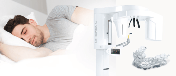
The first fully digital workflow for a more restful sleep
Easily identify patients who will be responders to oral appliance therapy and decrease time to therapy with a simple 3-step fully digital workflow.
The Orthophos SL 3D-Ai with MATRx plus, SICAT Air, and OPTISLEEP is a complete 3D solution for the analysis and appliance-based treatment of Obstructive Sleep Apnea (OSA).
This simple sleep solution integrates comparative airway data using the Orthophos SL 3D-Ai by Dentsply Sirona. Adding MATRx plus expands the SICAT Air and OPTISLEEP workflow resulting in a completely digital workflow from airway analysis, diagnostics of OSA, and oral appliance study, to the treatment of OSA with a custom-fitted oral appliance.

Mark your calendar for joint AAOMR/IADMFR meeting August 22-25, 2019
The American Academy of Oral & Maxillofacial Radiology and International Association
of Dento-Maxillo-Facial Radiology joint meeting will feature keynote speakers from world-renowned leaders and researchers in dental and medical radiology, diagnostic imaging workshops, and symposia covering the latest imaging technologies, modalities, and research in oral and maxillofacial radiology and dentistry. New methodologies and basis to clinical research in dental and medical radiology will also be covered.
This joint event will be the optimal setting for sharing the latest academic and research insights. The conference will be held at the Loews Philadelphia Hotel, Downtown Philadelphia August 22-August 25. Click here for more information and to register.
Full library. One price. 
ADA CE Online subscriptions are here. Get unlimited access to the entire ADA CE online library, including JADA, for one year from purchase. Access anywhere, anytime. With new courses being added every month, you’ll never run out of education opportunities. Group subscriptions are also available, check it out now.
Advertisement

The consulting editor for JADA+ Specialty Scan — Radiology is Laurie C. Carter, DDS, Ph.D., past president, American Academy of Oral and Maxillofacial Radiology. |
|
