
A band of keratinized mucosa (KM) around dental implants will improve patient esthetic satisfaction, according to a study published in the September issue of Journal of Periodontology.
Authors designed the study to measure the experiences and preferences of patients who had implants with and without KM and to assess patient satisfaction with the esthetics of peri-implant soft tissue.
A total of 24 patients received 28 2-stage dental implants: 17 were placed for molars, 9 for premolars, and 2 for anterior teeth. After second-stage surgery, 12 patients with 13 dental implants had KM on the buccal site (group 1), and 12 patients with 15 implants had no KM (group 2). Implants were restored with screw- or cement-retained fixed prostheses and evenly distributed across the dentition and between the arches.
The average patient age was 63.2 years for group 1 and 58.0 for group 2. More men than women (83.3%) comprised group 1, whereas the sex distribution was even for group 2. All 24 patients completed 3- and 6-month follow-up visits. At each visit, the examiner measured discomfort during toothbrushing and patient esthetic satisfaction using a 10-point visual analog scale (VAS).
Pretest results showed 93.3% and 73.3% agreement within patients for discomfort and esthetics scores, respectively. Authors measured the mean and standard error for the continuous variables KM and alveolar mucosa prospectively within group 1 and for the continuous variable recession (REC) within group 2. They calculated mean and standard deviation for continuous variables VAS for discomfort during toothbrushing and esthetic satisfaction and compared mucosal thickness (MT) and probing depth (PD) for groups 1 and 2. They conducted the Spearman correlation test to measure any correlation between MT, VAS for discomfort, VAS for esthetics, PD, REC, plaque index (PI), and bleeding on probing (BoP).
Authors found that patients without peri-implant KM were less satisfied with the esthetics of the soft tissue around the implant at the 3- and 6-month follow-up visits (P < .01). Esthetic satisfaction stayed high for implants with KM but decreased over time for those without KM. Implant location did not influence esthetics. The average PD and BoP after implant restoration were similar for groups 1 and 2. Authors noted no difference in plaque buildup between implants with and without KM or between implants that supported single- or multiunit restorations.
The width of KM in group 1 significantly increased after 6 months (P < .01). Patients reported greater esthetic satisfaction with a wider KM zone at 3 and 6 months (r, 0.633; r, 0.623, respectively). Authors noted an average REC of 0.27 millimeters after 3 months and 0.33 mm after 6 months in all implants. Lack of KM was not associated with patient discomfort during toothbrushing.
Read the original study here or contact the ADA Library & Archives for assistance.
Advertisement

AAP best evidence review: antimicrobial photodynamic therapy for the treatment of periodontitis and peri-implantitis
Antimicrobial photodynamic therapy (aPDT) as an adjunct to scaling and root planing (SRP) results in similar improvements in probing depth (PD) and clinical attachment level (CAL) as conventional periodontal therapy alone for patients with periodontitis and peri-implantitis. The findings are based on a systematic, best evidence consensus by the American Academy of Periodontology published in the July issue of Journal of Periodontology.
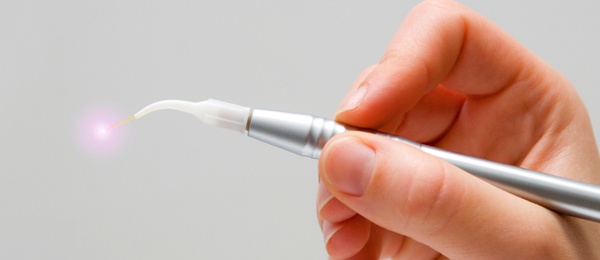
aPDT involves the use of a light-sensitive dye called a photosensitizer (PS) combined with visible light, stimulating the PS to form free radicals of oxygen which can have bactericidal properties. Authors conducted a review to measure the efficacy of aPDT in the nonsurgical and surgical treatment of patients with periodontitis and peri-implantitis. They included only randomized controlled trials (RCTs) with follow-up periods of at least 3 months. Eligibility criteria included treatment of patients with moderate to severe aggressive periodontitis (AgP) or chronic periodontitis (CP) (mean PD ≥ 5 millimeters) and measurement of mechanical root or implant surface debridement (scaling and root planing) with or without flap access versus aPDT as an adjunct to mechanical root debridement.
The search yielded 729 potential articles, of which 703 were excluded. Among the 26 included studies, 19 were conducted according to a split-mouth design and the remaining 7 according to a parallel design. A total of 69 patients with AgP, 567 patients with CP, and 50 patients with peri-implantitis were treated in the studies. Clinical outcome measures included change in PD, CAL, recession of gingival margin, bleeding on probing (BoP), bone defect fill, and microbial colonization and composition. Patient-centered outcomes included discomfort, esthetics, function, and treatment costs.
Authors used random-effects meta-analyses with continuous data and expressed pooled estimates as weighted mean differences (MDs) with 95% confidence intervals (CI). They measured discrepancies in estimates of treatment effects from different trials using the Cochran test for heterogeneity and the I2 statistic. They measured aPDT according to the type and phase of periodontal therapy: nonsurgical treatment of AgP and CP (4 RCTs), as part of basic procedures (13 RCTs), 3 months after basic procedures (3 RCTs), at least 1 year after regular periodontal maintenance (3 RCTs), nonsurgical treatment of patients with CP affected by risk factors such as smoking (1 RCT), and nonsurgical treatment of peri-implantitis (2 RCTs).
Only 1 study was considered to be at low risk of bias, 13 at high risk of bias, and 12 at unclear risk of bias.
All studies found significant intragroup improvements for CAL, PD, and BoP, but only 1 study found a superior mean PD reduction and mean CAL gain at 3-month follow-up for deep pockets (≥ 7 millimeters) when aPDT was combined with SRP. Two RCTs reported a reduction in the frequency of sites with moderate and deep (≥ 7 mm) pockets after SRP plus aPDT and SRP alone at 3-month follow-up. Two other studies found that aPDT produced greater reductions in bacteria levels than SRP alone.
Data from 3 trials showed a significantly greater reduction in PD for SRP plus aPDT than SRP alone (0.29 mm for sites with PD = 5-6 mm; 0.75 for sites with PD ≥ 7 mm; P < .05). Data from 13 other trials found that the use of SRP plus aPDT produced significant improvements in BoP, CAL, and PD, with no significant adverse effects. Pooled estimates from 11 trials comparing the use of SRP plus aPDT with SRP alone showed an additional reduction of 0.43 mm in mean PD for sites with PD of 5 through 6 mm. Pooled estimates on PD reduction and CAL gain (MD) at residual sites did not show statistically significant differences between SRP plus aPDT and SRP alone.
Read the original study here or contact the ADA Library & Archives for assistance.
AAP best evidence review: infrared lasers for the treatment of moderate to severe periodontitis
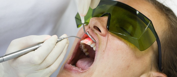
Scaling and root planing (SRP) plus infrared laser (diode, erbium:yttrium‐aluminum‐garnet, or neodymium:yttrium‐aluminum‐garnet (Nd:YAG) and erbium:yttrium‐aluminum‐garnet) or Nd:YAG laser alone can produce statistically significant gains in clinical attachment level (CAL) and probing depth (PD) in patients with moderate to severe aggressive periodontitis (AgP) or chronic periodontitis (CP). These gains were, however, relatively small (<1mm) as compared with SRP alone and the authors questioned the clinical relevance of this statistically significant difference. The findings are from a systematic, best evidence consensus by the American Academy of Periodontology published in the July issue of Journal of Periodontology.
Authors conducted the review to measure the effects of laser treatment, either alone or as an adjunct, to surgical and nonsurgical therapies on patients with periodontitis. They chose randomized controlled trials (RCTs) of at least 3 months that included adult patients 18 years or older with moderate to severe (mean PD ≥ 5 millimeters) or CP and RCTs that measured SRP with or without surgical flap access compared with infrared laser treatment alone or as an adjunct to mechanical root debridement.
Their search yielded 475 potential articles, of which 447 were excluded. The remaining 28 RCTs included in the review featured 25 conducted by split-mouth design and 3 conducted by parallel design. A total of 45 patients with AgP and 749 patients with CP were treated in the studies. Five studies included patient follow-up periods of at least 12 months. The remaining studies included follow-up periods of 3 through 6 months.
Clinical outcome measures included change (mean or percentage) in PD, CAL, recession of gingival margin (REC), bleeding on probing (BoP), bone defect fill, and microbial colonization or composition. Patient outcomes included discomfort, esthetics, function, and treatment costs.
Authors measured applications to infrared lasers according to the type and phase of periodontal therapy: nonsurgical treatment of AgP (2 RCTs), nonsurgical treatment of CP (15 RCTs), nonsurgical treatment of CP in patients after at least 1 year of regular periodontal maintenance (5 RCTs), nonsurgical treatment of CP in patients with risk factors such as smoking (2 RCTs), and surgical treatment of CP in patients with residual sites after basic procedures (4 RCTs).
Two trials found significant intragroup improvements to CAL, PD, and BoP. One of these 2 studies found that a 980-nanometer laser combined with SRP produced a superior mean PD reduction and mean CAL gain 6 months after treatment compared with either laser alone or SRP alone (P < .05). Both studies found that SRP plus laser therapies produced greater bacteria level reductions than SRP alone. Pooled estimated on PD reduction and CAL gain (MD) showed an additional reduction of 0.24 millimeters for sites treated with SRP plus diode infrared lasers.
Analysis of 15 other RCTs found that the use of SRP plus infrared lasers produced significant improvements in BoP, CAL, and PD. Pooled estimates on PD reduction and CAL gain (MD) showed addition PD reduction of 0.63 mm and CAL gain (MD) of 0.52 mm in sites treated with SRP plus diode laser.
Analysis of 5 other studies that measured the effect of laser therapy on the nonsurgical treatment of patients with CP and PD of at least 5 mm that underwent regular maintenance for at least 1 year found no improvements in PD or CAL. In an analysis of 4 other RCTs, only 1 reported significantly greater improvements in PD and CAP after flap access and laser debridement than with conventional open flap debridement at 6-, 12-, 24-, and 36-month examinations.
Read the original study here or contact the ADA Library & Archives for assistance.
AAP best evidence review: laser therapy for treatment of peri-implant mucositis and
peri-implantitis
Laser therapy combined with surgical and nonsurgical therapy to treat peri-implantitis provides minimal reduction of pocket depth (PD) and plaque index (PI), clinical attachment level (CAL) gain, and recession of gingival margin (REC). The findings are from a systematic review and meta-analysis published by the American Academy of Periodontology in the July issue of Journal of Periodontology. The review was able to form these conclusions only regarding erbium:yttrium‐aluminum‐garnet (Er:YAG), CO2 and diode lasers due to insufficient controlled evidence available for other lasers.
Authors designed the review to assess the potential for lasers to detoxify and treat peri-implant mucosis and peri-implantitis. The review assessed the use of lasers alone or as adjuncts in surgical or nonsurgical therapies. Primary outcomes included changes in PD, CAL, PI, REC, bleeding on probing (BoP), and marginal bone level (MBL).
Their search yielded 237 articles, of which 215 were excluded. Among the 22 chosen clinical trials, lasers were used as an adjunct to nonsurgical interventions in 13. Lasers were used surgically in 9 studies. A total of 11 studies (9 RCTs and 2 controlled clinical trials) were included for meta-analyses.
Most treated implants in the chosen studies were rough-surfaced implants. Smooth-surfaced implants were more commonly found in studies that featured a nonsurgical laser–treated approach. Studies that featured a surgical approach included a diode laser (3 studies), a carbon dioxide laser (2 studies), and the Er:YAG laser (4 studies). Studies that used a nonsurgical approach included a diode laser (8 studies) and Er:YAG laser (5 studies). Follow-up periods ranged from 6 through 60 months for studies that used a surgical approach and 6 through 12 months in studies that used a nonsurgical approach.
Studies that used a nonsurgical approach found weighted mean differences (WMDs) for PD reduction of 0.15 millimeters (95% CI, –0.28 to 0.57 mm; P = .50), CAL gain of –0.10 mm (95% CI, –0.30 to 0.10 mm; P = .32), BoP reduction of 21.08% (95% CI, 3.61% to 38.55%; P = .02), PI reduction of 0.07 (95% CI, –0.12 to –0.03; P = .002), MBL of –0.22 mm (95% CI, –0.43 to –0.01; P = .04), and REC of –0.11 mm (95% CI, –0.33 to 0.11 mm; P = .34).
Studies that used a surgical approach found WMDs for PD reduction of 0.45 mm (95% CI, –0.10 to 1.00 mm; P = 0.11), CAL gain of 0.22 mm (95 CI, –0.52 to 0.95 mm; P = 0.56), BoP reduction of 7.26% (95% CI, –38.77% to 53.29%; P = .76), and PI reduction of –0.09 (95% CI, –0.95 to 0.77; P = .84).
“Current evidence allowed for analysis of only Er:YAG, [carbon dioxide], and diode lasers,” the authors noted. “Studies on others failed to have controlled evidence to support their evaluation. Since the types of lasers analyzed have different modes of action, the limited number of included studies and patients/implants evaluated makes it difficult to warrant their therapeutic values.”
Read the original study here or contact the ADA Library & Archives for assistance.
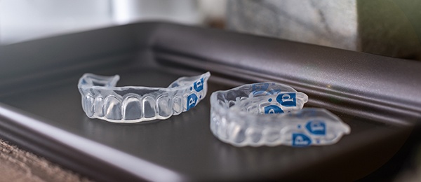
Perio Trays by Perio Protect
Prescription Perio Trays are innovative tools for adjunctive periodontal care. They have internal peripheral seals to deliver medication into shallow and deep pockets (>6mm), ideal for patients with generalized pocketing or struggling between maintenance visits. Research shows Perio Tray delivery of 1.7% hydrogen peroxide gel combined with SRP achieves better results than SRP alone. They are comfortable, convenient, easy-to-use. To learn more visit PerioProtect.com or Let'sTalkGumDisease.com.

AAP publishes updated periodontal, peri-implant disease classification 
The American Academy of Periodontology (AAP) has published the official proceedings from the 2017 World Workshop on the Classification of Periodontal and Peri-Implant Diseases and Conditions. These proceedings provide a comprehensive update to the previous disease classification established at the 1999 International Workshop for a Classification of Periodontal Diseases and Conditions.
Highlights from the 2017 proceedings include:
- Multi-dimensional staging and grading system for periodontitis classification (similar to an oncology model).
- Recategorization of various forms of periodontitis.
- Inaugural classification for peri-implant diseases and conditions.
The complete suite of review papers and consensus reports from the workshop, which was co-presented by the European Federation of Periodontology (EFP), is available in the June 2018 print and online issues of the Journal of Periodontology. Click here to read the proceedings, download practice resources, and check out the special behind-the-scenes “Making of the Workshop” documentary.
Free CE from the comfort of your own home 
ADA CE Online has a wide selection of on-demand courses that are free for all, including this course by Dr. Sebastian Cianco, Baking Soda Dentifrices: Benefits for Oral Health. Benefit your patients’ smiles by learning all about the clinical, esthetic, and antimicrobial benefits of baking soda.
This course is sponsored by Arm & Hammer.
Advertisement

The consulting editor for JADA+ Specialty Scan — Periodontics is Tapan Koticha, BDS; Diplomate, American Board of Periodontology, Director, Graduate Periodontics, University of Oklahoma Health Sciences Center, College of Dentistry |
|
