
Maxillary transverse dimensions in patients with and without impacted canines
Impacted maxillary canines are a common finding in orthodontics, and cone-beam computed tomography (CBCT) is an effective and efficient diagnostic and treatment planning technique. The purpose of this retrospective study was to compare the maxillary transverse dimensions of patients with impacted maxillary canines with those of patients without canine impaction. The study was published in the October 2018 issue of American Journal of Orthodontics & Dentofacial Orthopedics.
The study sample consisted of 86 patients with at least 1 impacted maxillary canine (45 unilateral [20 labial, 25 palatal] and 41 bilateral [21 labial, 7 palatal, 13 combined labial and palatal) and 67 patients with no impacted maxillary canines (control group) from 2 private diagnostic centers in Lima, Peru. Participants in the 3 groups had similar vertical growth patterns, skeletal sagittal relationships, and maxillary sagittal positions.
According to the selection criteria, participants were required to have no more than 2 millimeters of anterior dental crowding (measured between the central and lateral incisors). In addition, researchers excluded participants with a history of orthodontic treatment and those with cleft lip or palate, craniofacial anomalies, head and neck syndromes, tumors, trauma or history of trauma, missing teeth or dental agenesis, or other maxillary lesions.
CBCT scans were acquired, and the researchers obtained CBCT synthesized cephalograms to match the groups according to vertical and sagittal characteristics.
For maxillary width measurements, the authors measured maxillary transverse dimensions at 4 levels: first molar basal width (MBW), first molar alveolar width (MAW), first premolar basal width (PMBW), and first premolar alveolar width (PMAW).
The study findings showed statistically significantly greater MBW (P = .030), MAW (P < .001), and PMAW measurements (P < .001) in the control group than in the groups with unilateral and bilateral impacted maxillary canines. The researchers did not observe any significant differences in PMBW measurements among the 3 groups (P = .063). In addition, they did not observe any significant differences in transverse measurements between the 2 groups with impacted canines.
In this retrospective study, patients with unilateral or bilateral impacted maxillary canines had smaller transverse measurements than patients without impacted maxillary canines. Longitudinal studies are needed to determine whether canine impaction is secondary to reduced maxillary width, the authors wrote. Moreover, clinicians should consider the relationship between maxillary width and canine impaction during diagnosis and treatment planning, the authors concluded.
Read the original article here or contact the ADA Library & Archives for assistance.
Advertisement

Canine eruption after orthopedic arch expansion
The aim of this prospective, longitudinal clinical trial was to assess the short-term impact of rapid maxillary orthopedic expansion (RME) on the eruption paths of ectopically erupting canines (EECs) and normally erupting canines (NECs) in the mixed dentition. The study was published in the October 2018 issue of American Journal of Orthodontics & Dentofacial Orthopedics.
Thirty-two patients (13 boys, 19 girls; mean age, 9.53 years) with 49 EECs made up the first treatment group. A second treatment group (8 boys, 10 girls; mean age, 9.25 years) was composed of 9 patients from the EEC group who had unilateral NECs and 9 patients with bilateral NECs, for a total of 27 NECs.
Inclusion criteria were patients with mixed dentition, RME indicated as a result of transverse maxillary deficiency, biological maturity corresponding to one-half through two-thirds of the canine root completed, ectopic eruption of at least 1 maxillary canine overlapping a portion or all of the lateral incisor root, no previous orthodontic treatment, no supernumerary teeth or lateral incisor agenesis, no early loss of primary teeth that could benefit canine eruption, no oral pathology associated with eruption disturbance or a history of dental trauma, and no craniofacial anomaly or syndrome.
In addition to the 2 treatment groups, an untreated control (UC) group was included, which was composed of 54 normally erupting maxillary canines in 36 participants (17 boys, 19 girls; mean age, 9.03 years) who had been followed longitudinally during growth.
The researchers obtained panoramic radiographs before expansion (T1) and 1 year after expansion (T2). One investigator digitized the radiographs, and a second investigator reviewed them for landmark identification.
The study findings showed that canines in the 2 RME treatment groups achieved greater angular, vertical, and horizontal changes toward a more normal position than did those in the UC group.
At T1, canines in the EEC group were substantially more ectopic than those in the UC and NEC groups. After RME, 22 of 49 canines (44.9%) in the EEC group migrated to no longer overlap the lateral incisor, and only 2 still completely overlapped the lateral incisor root, as seen on the panoramic radiograph.
Regarding vertical changes, 27 canines (55.1%) in the EEC group and 18 (66.7%) in the NEC group were at the occlusal level after RME, compared with 21 canines (38.9%) in the UC group.
The treatment changes with RME had a favorable impact on the eruption path of ectopic and normally erupting canines, the authors wrote. In addition, the positions of teeth adjacent to the canines were significantly affected by RME, and these changes also might be associated with improvement in the position of the EEC.
Based on the results of this study and others, the researchers advised clinicians to consider RME in patients who have constricted arch form and in whom the potential for canine impaction is mild. For similar patients in whom the potential for canine impaction is moderate to severe, clinicians should consider extraction of the primary canine along with RME, the authors wrote.
Read the original article here or contact the ADA Library & Archives for assistance.
Advertisement

Evaluating changes in normal occlusion over a 40-year period
The literature contains little information regarding normal occlusal changes through the sixth decade of life. In this observational study, researchers evaluated the changes in dental arch dimensions, tooth size, and incisor crowding in participants with normal occlusion over a 40-year period. The study was published in the August 2018 issue of American Journal of Orthodontics & Dentofacial Orthopedics.
The initial study sample comprised 82 white Brazilians (39 men, 43 women) recruited from 1967 through 1974. At age 13 years (T1), all participants had a clinically acceptable occlusion in the complete permanent dentition, dental and skeletal Class I relationships, no crossbites, normal overjet and overbite, and no more than 2 mm of incisor crowding, with no previous orthodontic treatment. At T1 and at T2 (17 years of age), researchers obtained dental models and cephalometric radiographs.
From April 2015 through May 2016, the researchers recalled participants (60 years of age) and obtained dental models (T3). They excluded participants who had undergone orthodontic treatment from T1 to T3, had complete tooth loss, or had no dental models from any of the 3 time points. The final sample was composed of 22 participants (12 men, 10 women).
The dental models were digitized with a 3-dimensional scanner (R700, 3Shape), and measurements were obtained using 3-dimensional software (OrthoAnalyzer, 3Shape). Measurements included tooth size, clinical crown height, arch width, arch perimeter, arch length, palatal depth, incisor crowding index, overjet, overbite, and curve of Spee.
One operator performed all measurements. She measured one-half of the sample twice, with an interval of at least 1 month. The study results showed excellent intrarater agreement, with intraclass correlation coefficients ranging from 0.78 through 0.99.
According to the study findings, the aging process affected most variables from T1 to T3. Mesiodistal tooth size, mandibular intercanine width, arch length, arch perimeter, overbite, and curve of Spee decreased, while clinical crown height, incisor crowding, and palatal depth increased. Overjet and maxillary arch widths remained stable over time, the authors wrote.
The researchers did not observe any changes in mesiodistal tooth size from T1 to T2, but the clinical crown height of most teeth increased. Maxillary arch length and perimeter, overbite, and curve of Spee decreased during this period, but mandibular crowding and palatal depth increased from T1 to T2.
From T2 to T3, mesiodistal tooth size, mandibular intercanine width, arch length, arch perimeter, and overbite decreased. The change in anterior overbite may be due to mandibular growth and tooth wear, with men showing a greater decrease than women. However, the clinical crown height of most posterior teeth and anterior crowding increased during this period.
One study limitation was the finding that at T3 many teeth (mean, 3.09) had been lost. This rate of tooth loss, which was higher than that in previous studies, was probably the result of the long follow-up period, older age (60 years) at the final evaluation, and lack of water fluoridation in the city until 1975, the authors wrote.
The researchers noted that increases in clinical crown height and dental crowding and decreases in mesiodistal tooth size, arch length, arch perimeter, and overbite might be expected with aging in people who have normal occlusion. They urged clinicians to be more conservative in recommending periodontal procedures for surgical crown lengthening because of the natural increase in clinical crown height over time. In addition, the authors advised clinicians to consider procedures that minimize incisor wear in adults.
Future studies are needed to compare the aging changes of people with normal occlusion with those of people who have undergone occlusion-related orthodontic treatment, the authors concluded.
Read the original article here or contact the ADA Library & Archives for assistance.
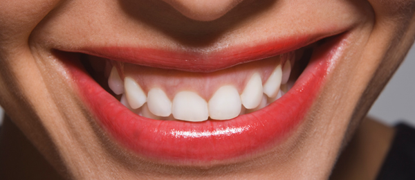
Laypeople’s perceptions of altered gingival characteristics on smile esthetics
Many studies have investigated laypeople’s perceptions regarding altered dental esthetics, but few have focused on soft-tissue factors. In this study, published in the July 2018 issue of American Journal of Orthodontics & Dentofacial Orthopedics, researchers evaluated laypeople’s perceptions of altered gingival characteristics. They wanted to know, in the context of a smile, what are the least and the most noticed gingival characteristics, as well as the level of deviation at which these attributes become detrimental in laypeople’s perceptions.
The researchers selected a highly attractive frontal close-up smile of a 22-year-old woman to serve as the study control. They cropped the image to show only the lips, nasal tip, and mentolabial fold. They then altered the image to create 65 photographs with manipulated soft-tissue features. The investigators also included 2 unaltered photographs of the ideal smile to check for reproducibility. The images were then printed and placed randomly in a photo album.
The following modifications were made to the image of the ideal smile:
- discrepancies in the position of the free gingival margin without recession;
- variations in the positions of interdental papilla (black triangles);
- discrepancies in the position of zenith;
- color changes caused by varying degrees of gingival inflammation;
- color changes caused by varying degrees of pigmented gingiva;
- discrepancies in the position of the free gingival margin with recession;
- variations in gingival contours.
The researchers altered each of these 7 factors with progressive variations—both unilateral and bilateral—in the original control photograph.
The randomly selected lay evaluators consisted of 45 men and 55 women (age range, 18-40 years). The researchers asked them to rate the attractiveness of the 67 images using a forced distribution Q sort method. According to this method, evaluators were required to place a defined number of photographs in each of 13 columns in a table according to their personal esthetic preferences. The columns were labeled from –6 (most unattractive) to +6 (most attractive). Evaluators took an average of 15 to 20 minutes to complete the survey.
The lay evaluators rated the ideal photograph (control) highest in attractiveness, with a mean (standard deviation [SD]) ranking of 3.93 (1.74). Alterations in the position of gingival zenith were the next-highest rated factor (mean [SD] ranking, 1.87 [1.33]) followed closely by variations in gingival contours (mean [SD] ranking, 1.81 [1.34]). Discrepancies in the position of the free gingival margin without recession received a mean (SD) ranking of 0.44 (1.39) by evaluators, while discrepancies in the position of the free gingival margin with recession received a mean (SD) ranking of –1.17 (1.20). Evaluators gave a mean (SD) ranking of –1.44 (2.53) to color changes caused by varying degrees of gingival inflammation, followed by a mean (SD) ranking of –2.54 (1.87) to color changes caused by varying degrees of pigmented gingiva. They gave the most negative ranking to variations in the position of interdental papilla (black triangles) (mean [SD] ranking, –2.60 [1.78]).
The lay evaluators also generally rated unilateral or asymmetric alterations in the smile image as more unesthetic than bilateral or generalized alterations.
The authors noted that clinicians should focus on the gingival factors that are most important, as well as on how these characteristics are perceived by patients. The results of this study indicate that when designing a smile, achieving “ideal” esthetic gingival parameters is not essential, but differences, especially in symmetry, can adversely affect laypeople’s perceptions. Future studies can compare the laypeople’s gingival characteristic perceptions with those of dentists, which will allow clinicians to develop treatment plans that are better balanced between patient and professional esthetic goals, the authors concluded.
Read the original article here or contact the ADA Library & Archives for assistance.

The SunClear advantage
More and more patients are asking for aligners but, due to costly aligner lab fees, many are forced to decline treatment or go online (for do-it-yourself aligners). Thanks to the SunClear 3D aligner design software and manufacturing solution, digital orthodontics has now become more affordable. SunClear aligners are the clear alternative to expensive and overpriced aligner systems.
• Save up to 50% on aligner lab fees.
• High-quality aligners design ready for approval ready in a matter of days after the impression has been received by the lab.
AAO Annual Session is May 3-7 ![]() The Los Angeles Convention Center in Los Angeles, California is the site of the American Association of Orthodontists’ 2019 Annual Session May 3 through May 7. This year’s meeting showcases rich learning experiences in biomechanics, CBCT technology, sleep apnea, Class II and Class III treatment, management of adult patients and more, along with practice management lectures and learning geared toward clinical and administrative staff teams. Special events and the world’s largest exhibit hall of products and services that are essential to contemporary orthodontic practices round out the AAO experience. Find details on the 2019 AAO Annual Session and register online here.
The Los Angeles Convention Center in Los Angeles, California is the site of the American Association of Orthodontists’ 2019 Annual Session May 3 through May 7. This year’s meeting showcases rich learning experiences in biomechanics, CBCT technology, sleep apnea, Class II and Class III treatment, management of adult patients and more, along with practice management lectures and learning geared toward clinical and administrative staff teams. Special events and the world’s largest exhibit hall of products and services that are essential to contemporary orthodontic practices round out the AAO experience. Find details on the 2019 AAO Annual Session and register online here.
AAO offers orthodontics resource for parents![]() An online primer for parents, The Parents’ Guide to Orthodontics, is offered by the American Association of Orthodontists. It walks readers through orthodontic treatment in an easy-to-digest Q&A format, from the first suspicion that treatment might be ahead to what to expect post-treatment. The guide includes an orthodontic glossary written for the lay person.
An online primer for parents, The Parents’ Guide to Orthodontics, is offered by the American Association of Orthodontists. It walks readers through orthodontic treatment in an easy-to-digest Q&A format, from the first suspicion that treatment might be ahead to what to expect post-treatment. The guide includes an orthodontic glossary written for the lay person.
Live patient Botox and Dermal Fillers courses at ADA Headquarters  Join us May 10-11, 2019 at ADA Headquarters in Chicago, IL for Botulinum Toxins and Dermal Fillers for Every Dental Practice. Now’s your chance to learn Botox and Dermal Filler treatment techniques that you will be able to immediately implement into your practice, led by Dr. Louis Malcmacher and his team at the American Academy of Facial Esthetics. Register now!
![Dr. Graber Preferred Photo[2]](https://pages.ada.org/hs-fs/hubfs/Dr.%20Graber%20Preferred%20Photo%5B2%5D.jpg?width=140&name=Dr.%20Graber%20Preferred%20Photo%5B2%5D.jpg)
The consulting editor for JADA+ Specialty Scan — Orthodontics is |
|
