
Is chronic mucosal trauma linked to oral squamous cell carcinoma?
Chronic trauma has been suggested as an etiologic risk factor for oral squamous cell carcinoma. In this study, researchers investigated whether inflammatory fibrous hyperplasia associated with removable dental prostheses was linked to higher rates of loss of heterozygosity (LOH) than normal oral mucosa. The research was published online March 8 in Journal of Oral Pathology & Medicine.
The researchers selected 15 formalin‐fixed paraffin‐embedded (FFPE) samples of inflammatory fibrous hyperplasia associated with removable dental prostheses, and 2 pathologists confirmed the diagnosis. In addition, they included 7 normal oral mucosa FFPE samples (control group) obtained during biopsies of nonneoplastic oral lesions.
The investigators manually microdissected epithelial and connective tissues and using a QIAamp DNA FFPE Tissue Kit (Qiagen, Hilden) isolated genomic DNA. They used 4 polymorphic microsatellite markers—D3S1300 at 3p14.2, D9S1748 at 9p21, D17S974 at 17p13, and D17S1289 at 17p12—to investigate LOH, an early genetic alteration in oral tumorigenesis. The researchers used capillary electrophoresis to conduct the LOH analyses.
Of the 15 inflammatory fibrous hyperplasia samples, 2 (13%) experienced LOH at markers D17S974 and D9S1748, the authors wrote. The frequency of allelic loss in the inflammatory fibrous hyperplasia group was 16.6% at D17S974 and 11.1% at D9S1748. None of the oral mucosa samples in the control group exhibited LOH.
In light of the study findings, the authors concluded that inflammatory fibrous hyperplasia associated with removable dental prostheses does not have the potential for malignant transformation.
Read the original article or contact the ADA Library & Archives for assistance.
Advertisement

The prevalence of oral mucosal disorders during pregnancy
Pregnant women may be at higher risk of developing oral disorders, primarily as a result of immune and hormonal changes. In this systematic review and meta-analysis, researchers assessed the prevalence of oral mucosal disorders during pregnancy. The study was published in the April issue of Journal of Oral Pathology & Medicine.
Researchers conducted an electronic search of the Cumulative Index to Nursing and Allied Health Literature , Latin American and Caribbean Health Sciences Literature, LIVIVO, PubMed, Scopus, Web of Science, Google Scholar, OpenGrey, and ProQuest databases. They also searched the gray literature and conducted a manual search of reference lists in included studies. Two authors independently selected studies based on a review of article titles and abstracts.
The inclusion criteria consisted of observational studies investigating the prevalence of oral mucosal disorders in pregnant women without language or time restrictions. Included disorders were aphthous ulceration, benign migratory glossitis, candidiasis, cheilitis, gingival epulis, herpes-associated lesions, leukoedema, lichen planus, papilloma, pemphigus, pemphigoid, pyogenic granuloma, and ulcers. The researchers did not assess gingivitis and periodontitis because systematic reviews and meta-analyses have been published on this topic.
The authors excluded studies that included participants with systemic chronic diseases, studies that included participants receiving treatment with medications potentially associated with oral mucosal disorders, and studies that included smokers or people who consumed alcohol on a long-term basis.
The 2 reviewers retrieved 3,508 articles from the databases. After duplicates were removed, a total of 1,833 articles remained. Of these, 1,802 were excluded after a comprehensive review of article titles and abstracts. The 2 reviewers then conducted a full-text assessment of 31 studies, which resulted in 15 studies meeting the eligibility requirements for the systematic review and meta-analysis.
A total of 5,935 participants were included in the meta-analysis, with sample sizes ranging from 86 through 1,600 people. Participants’ ages ranged from 10 through 50 years. All studies were published from 1954 through 2017 and conducted in 9 countries (Egypt, Ghana, India, Iran, Mexico, Nigeria, Pakistan, Turkey, and Venezuela).
The reviewers used the Joanna Briggs Institute Critical Appraisal Checklist for Studies Reporting Prevalence Data to evaluate risk of bias in the selected studies. In addition, they assessed the overall quality of evidence by means of the Grading of Recommendations Assessment, Development and Evaluation (GRADE) criteria.
The overall prevalence rate (PR) of oral mucosal lesions was 11.8% (I², 98%; 95% confidence interval [CI], 6.5% to 18.4%), the authors wrote. The most commonly reported lesions were hyperplasia (PR, 17.1%) (I², 97%; 95% CI, 5.9 to 32.7), morsicatio buccarum (PR, 9.9%; I², 96%; 95% CI, 1.3 to 25.2), oral candidiasis (PR, 4.4%) (I², 92%; 95% CI, 0.5 to 11.8), pyogenic granuloma (PR, 3%) (I², 90%; 95% CI, 1.6% to 4.8%), and benign migratory glossitis (PR, 2.8%) (I², 0.0%; 95% CI, 1.9 to 3.8).
The researchers found a moderate risk of bias in 9 studies and a low risk in 6 studies. Using the GRADE criteria, they determined that the quality of evidence in the 15 studies was very low. They attributed this finding to small sample sizes, wide confidence intervals, substantial heterogeneity of diagnostic methods, and study designs that did not allow investigators to evaluate participants according to trimester.
Future studies should include adequate sample sizes and use more homogeneous methods and standardized diagnostic criteria, the authors concluded.
Read the original article or contact the ADA Library & Archives for assistance.
Advertisement
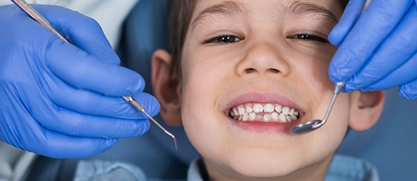
The oral health status of children who are long-term cancer survivors
In recent years, more attention has been focused on the long-term adverse effects of cancer treatments in children. The objective of a study published in the May issue of Supportive Care in Cancer was to evaluate the oral health status and craniofacial development of children who were long-term survivors of malignancies.
The study group was composed of 53 children (41 boys, 12 girls) with a mean age of 10 years, 4 months, who were referred from the Department of Pediatric Oncology at Gazi University in Ankara, Turkey. The mean time since cessation of cancer treatment was 2 years, 4 months (range, 1-5 years). The control group was made up of 40 healthy children (19 boys, 21 girls) with a mean age of 12 years, 4 months, who were recruited from the Department of Pediatric Dentistry.
An experienced periodontist conducted the oral examinations in both the test and control groups. The eruption status of permanent teeth and any crown anomalies or soft-tissue abnormalities were recorded. The clinician also evaluated participants’ periodontal status and oral hygiene using the gingival index and plaque index. In addition, the periodontist evaluated the children for caries and recorded the decayed, missing, and filled teeth index scores for both primary (dmft) and permanent (DMFT) dentition. All children in both groups were in mixed dentition.
Thirty-one of the 53 children in the study group and 26 of the 40 children in the control group underwent maxillofacial radiography for evaluation of craniofacial development. Panoramic, lateral cephalometric, and hand-wrist radiographs were obtained in these participants.
The researchers observed enamel discoloration in 30 children (255 teeth) in the test group and in 9 children (72 teeth) in the control group. The differences between the groups were statistically significant at both the patient (P < .001) and tooth (P < .048) levels, the authors wrote. The study findings also showed enamel hypoplasia in 32 patients (147 teeth) in the study group and in 17 patients (62 teeth) in the control group. This difference was statistically significant at the tooth level (P < .004) but not at the patient level (P = .131).
Root malformations were observed in 22 of 53 children (49 teeth) in the study group and 7 of 40 children (11 teeth) in the control group. The differences between the 2 groups were statistically significant only at the patient level (P < .013).
The gingival index and plaque index scores were significantly higher in children in the study group than in those in the control group. However, the mean dmft and DMFT scores in the study and control groups were not significantly different, the authors wrote.
Regarding craniofacial development, the radiographic examinations revealed no statistically significant differences between children in the study and control groups.
The results of this study showed that antineoplastic therapy or childhood cancer resulted in a higher prevalence of various tooth malformations. Children treated at younger ages experienced more severe defects, suggesting that immature teeth are at greater risk of experiencing developmental disturbances than fully developed teeth. However, future studies should include larger sample sizes and longer follow-up periods.
Early diagnosis, treatment, and individualized prevention programs based on plaque control are essential for minimizing the oral consequences of cancer treatment and improving oral health, the authors concluded.
Read the original article or contact the ADA Library & Archives for assistance.

The impact of oral health care motivation on patients with oral lichen planus
The role of dental plaque in gingival lesions resulting from oral lichen planus (OLP) has not been fully delineated. In this longitudinal study, published online April 4 in Oral Diseases, researchers examined the effect of home-based oral health care motivation on quality of life and clinical parameters in patients with OLP-associated gingival lesions.
Investigators at the Hospital of the University of Parma in Parma, Italy, randomly assigned 60 patients (11 men, 49 women) with a biopsy-proven diagnosis of OLP to an intervention (n = 29) or a control (n = 31) group. Those in the intervention group took part in a 30-minute motivational session during which a dental hygienist instructed them on use of atraumatic procedures to remove bacterial biofilm from buccolingual and proximal dental surfaces. Patients in the intervention group also received 2 manual toothbrushes and dental picks with soft rubber bristles and flexible plastic stems. Participants in the control group did not receive any instructions and were asked to maintain their normal oral hygiene habits.
The primary outcome variable was quality of life, as assessed by the Oral Health Impact Profile (OHIP‐14). The secondary outcome variables were maxillary and mandibular gingival pain, as indicated on a visual analog scale; a modified Quigley Hein plaque index; and clinical disease severity (according to a modified Escudier index). The researchers assessed these variables at 0, 4, and 20 weeks and analyzed the results using an analysis of variance model for factorial design.
All patients were followed up at 4 weeks and 20 weeks, the authors wrote. At the 4-week visit, the same dental hygienist reinforced the oral hygiene instructions given at baseline to participants in the intervention group. (At the end of the study, control group participants also received the oral hygiene instructions.) In addition, 1 trained and calibrated operator recorded all data at baseline, 4 weeks, and 20 weeks.
At baseline, the researchers observed no significant differences between the intervention and control groups with regard to any variable. However, at 4 weeks, the plaque index, lesion severity score, and lesion activity score (lesion site score × lesion severity score) were significantly lower in the intervention group than in the control group (P < .05). Regression analysis revealed a significant positive trend for OHIP-14 in the intervention group compared with the control group (P < .05). These differences were maintained at 20 weeks. Pain did not differ significantly in the 2 groups (P = .408).
In this randomized controlled trial, the clinical severity of gingival lesions and OHIP-14 improved with plaque control, the authors wrote. They stressed the need for dental hygienists to be involved in the multidisciplinary management of patients with OLP and symptomatic gingival lesions.
Read the original article or contact the ADA Library & Archives for assistance.
2019 AAOMP Annual Meeting
Join the American Academy of Oral & Maxillofacial Pathology at its Annual Meeting, June 7 - 12, in Miami, FL.
The annual AAOMP Seminar panel session, moderated by Dr. Indraneel Bhattacharyya, Division Head, University of Florida Oral and Maxillofacial Pathology, will showcase interesting cases from the University of Florida Oral Pathology team.
Earn additional CE credits by attending the following sessions:
- Symposium — “HPV & Oro-pharyngeal and Oral Cancer” — Dr. Gypsyamber D’Souza, Associate Professor Epidemiology at the John Hopkins Bloomberg School of Public Health.
- Slide seminar — “Head and Neck Pathology” — Dr. Bruce Wenig Section Head of Anatomic Pathology External Network Consultation Services and Head and Neck–Endocrine Pathology Program Moffitt Cancer Hospital.
- Slide seminar — “Hematopathology: Lymphoid Lesions of the Head and Neck” — Dr. L. Jeffrey Medeiros, Chief of the Lymphoma Section in the Department of Hematopathology at the University of Texas M.D. Anderson Cancer Center.
- Founders Seminar — “Newly Defined (and Recently Refined) Head and Neck Tumors” — Dr. Justin Bishop, Associate Professor of Pathology, Otolaryngology-Head and Neck Surgery, and Oncology at Johns Hopkins Hospital.
- Gorlin Lecture — “Genetic Disorders Involving the Oral and Maxillofacial Region: A Dermatologist’s View” — Dr. Rudolph Happle, world-renowned dermatologist, Professor Emeritus at the Department of Dermatology, University of Marburg ny and Professor Emeritus at the Department of Dermatology, Philipp University.
For more information or to register, visit the meeting website.
Pathology Track at ADA FDI World Dental Congress! The Pathology Track at ADA FDI 2019 is just one of many disciplines offering you critical educational content on some of the most important topics facing dentistry today. Join us Sept. 4-8 in San Francisco, California, and select a pathology course that speaks to your personal needs and interests.
Most of these CE courses are priced at just $25, $50, or $75.
Tracks to consider, among many others:
- Mouthful of Lesions in Peewees: Maximizing Infant Oral Health (#5140), CE HOURS: 2.5
- Come in and Catch It: The Review That Sticks (#6140), CE HOURS: 2.5
- Come in and Catch It: The Radiology Review (#6141), CE HOURS: 2.5
- I Was on the Internet Last Night (#7142), CE HOURS: 3
- Trauma Management (#6139), CE HOURS: 2.5
Register today at ADA.org/meeting.
ADA CE Online pathology courses  Need CE? ADA CE Online has hundreds of hours of CE that you can earn from the comfort of your own home. Take a look at our courses focused on pathology that you can implement in your own practice. Too many to choose from? Take them all! Get an ADA CE Online subscription for one year and enjoy unlimited access. Subscribe now!

The consulting editor for JADA+ Specialty Scan — Oral Pathology is Faizan Alawi, DDS, Associate Dean for Academic Affairs and Associate Professor of Pathology, School of Dental Medicine; Associate Professor of Dermatology, Perelman School of Medicine; Director, Penn Oral Pathology Services; University of Pennsylvania |

The associate consulting editor for JADA+ Specialty Scan — Oral Pathology is Bruno C. Jham, DDS, MS, PhD; Associate Professor and Associate Dean for Academic Affairs; College of Dental Medicine – Illinois, Midwestern University |
|
