
Overbite, overjet influence soft-tissue profile in mature adults
A profile photograph offers “significantly relevant” information about occlusal relationships of anterior teeth in patients with negative overjet. The severity of negative overjet also increases the severity of mandibular prognathism. The findings are from a study published in the January issue of American Journal of Orthodontics & Dentofacial Orthopedics.
Authors designed the study to measure the association between overjet, overbite, and soft-tissue profile characteristics in a group of middle-aged patients. The study population included 1,630, 46-year-old adults (712 men, 919 women) from the Northern Finland Birth Cohort of 1966. Less than 20% of the study population had undergone previous orthodontic treatment.
The standardized clinical examination for all patients included intraoral occlusal measurements, bite registration, and clinical photographs. Images consisted of 1 profile and 2 frontal photographs (resting and posed smiling). Authors noted that detailed measurements for overjet and overbite had been published previously.
Authors identified 11 soft-tissue facial landmarks for their analyses. They used independent sample t tests to measure profile differences between men and women and linear regression models to measure the associations among angular and linear profile measurements, overjet, and overbite.
Authors found that the regression model explained roughly 30% of the variability in overjet and about 22% of the variability in overbite. They found that overjet was related more significantly to upper and lower anteroposterior lip position and to upper and lower facial height. Overbite was more strongly associated with anteroposterior position of the lower lip, pogonion, and soft-tissue B point. The anteroposterior position of the upper lip was more strongly associated with overjet than other factors when authors examined independent variables.
A total of 1,313 patients (80.55%) had an overjet 1 through 4 millimeters, and 1,158 (71.04%) had an overbite 1 through 4 mm. Authors also found that facial soft-tissue measurements could not accurately predict the severity of the malocclusion in most subgroups.
The authors found moderate correlations between facial characteristics and severe positive overjet, deep bite, and open bite. In the negative overjet subgroup, facial soft-tissue measurements predicted 44.2% of the variation in overjet value, indicating a strong association between facial morphology and negative overjet.
“Soft tissue A-point, soft tissue B-point and the upper and lower lips appeared to statistically influence the results of the regression model more than other measurements,” authors concluded. “It may be presumed that they relate better to underlying skeletal differences between subjects. To create predictive models and improve the diagnostic value of facial photographs, measurements including these structures could be beneficial.
Read the original study here or contact the ADA Library & Archives for more help.
Advertisement
Walking bleach technique with hydrogen, carbamide peroxides for tooth whitening
Dentists who use the walking bleach technique to whiten teeth can achieve a high level of color matching with minimal rebound, along with a positive effect on patients’ psychosocial impact. The study, published in the January/February issue of Operative Dentistry, found that 35% hydrogen peroxide and 37% carbamide peroxide were highly effective for the nonvital tooth walking bleach technique.
Authors designed the randomized, double-blind study with a 2-fold purpose:
- to measure the amount of color rebound in patients who received bleaching of nonvital teeth with 35% hydrogen peroxide or 37% carbamide peroxide using the walking bleach technique;
- to measure the impact on esthetic self-perception and psychosocial impact.
Authors evenly split a group of 50 patients according to bleaching agent. For the preparation session, authors prepared the canal under absolute isolation, removing 3 millimeters of endodontic filling from the cementoenamel junction (CEJ). They then applied and cured a 2-mm seal of resin-reinforced glass ionomer to protect the endodontic treatment. Authors limited the seal’s coronal limit to 1 mm below the CEJ to ensure whitening.
For the next 4 whitening sessions, authors applied the whitening agent according to the manufacturer’s instructions using the walking bleach technique: gel placed in the cavity pulp chamber in the presence of mild moisture. They used temporary cement to close the cavity until the next session 7 days later. The procedure was repeated in each session for up to 4 weeks. Authors used the same amount of gel in each session.
For the final session, authors washed the cavity access, then inserted a temporary restoration for 7 days before the final restoration with composite resin was finished.
Two calibrated examiners measured tooth color at baseline and again at 1 week, 1 month, 6 months, and 1 year after bleaching. Authors arranged color guides ranging from highest (B1) to lowest (C4) and measured color changes using Vita Classical Shade units (DELTASGUs).
Authors administered the Oral Health Impact Profile questionnaire at 1 week, 1 month, 6 months, and 1 year after bleaching to measure the impact on patient quality of life. The Psychosocial Impact of Dental Aesthetics Questionnaire (PIDAQ) grouped 23 questions into 4 impact areas: dental self-esteem, social impact, psychosocial impact, and esthetic concerns. Authors used the Wilcoxon test to compare the effect of color treatment with each group and the Mann-Whitney test for comparisons between groups.
Authors found that color changes (DELTAE [standard deviation]) 1 year after treatment with the walking bleach technique were 15.11 (5.59) for hydrogen peroxide and 13.17 (5.13) for carbamide peroxide. The PIDAQ score was significantly different at the start of the study compared with a year later for all factors except esthetic concerns for the carbamide peroxide group. Authors also found statistically significant differences in the carbamide peroxide group (P < .05) for total score, functional limitations, and psychological disabilities but no statistically significant differences between the 2 groups at the 1-year follow-up.
Read the original article here or contact the ADA Library & Archives for help.
OHRQoL of preschoolers and families after SDF treatment
The oral health–related quality of life (OHRQoL) of preschool-aged children is not affected by silver diamine fluoride (SDF) therapy at 6 months, while parents whose children have higher caries experience more parental distress following SDF treatment. The findings are from a study published in the February issue of Journal of Dentistry.
Authors designed the study to measure changes in OHRQoL of preschool-aged children from 6 kindergartens in Hong Kong who received SDF treatment for dental caries in a school oral health program. Researchers conducted a clinical oral examination at baseline and again at 6 months.
All children were examined lying down. Authors used the decayed, missing and filled teeth index (dmft) to record caries status. Carious lesions were explored with a Community Periodontal Index probe. Authors classified a carious lesion as active if gentle probing detected softness or as arrested if the dentin surface was hard to probe. They defined the caries arrest rate as the number of arrested SDF-treated surfaces at 6 months divided by the number of active caries lesions receiving SDF at baseline. They used the visible plaque index to measure oral hygiene status.
Authors administered a Chinese version of the Early Childhood Oral Health Impact Scale (ECOHIS) questionnaire at baseline and again at 6 months to measure OHRQoL. ECOHIS features 13 items grouped into a child impact section and a parent impact section. Authors used a 5-point coded scale for responses ranging from 0 (never) through 4 (very often). Total scores ranged from 0 through 52.
A total of 117 children were treated with 38% SDF in a school-based setting. Their mean (standard deviation [SD]) dmft score at baseline was 4.9 (3.8). Most children (69.9%) had 1 through 5 carious teeth. A total of 113 returned a completed questionnaire at 6 months. Their mean age was 4.6 years. The mean (SD) number of active caries teeth receiving SDF therapy at baseline was 4.6 (3.6). Most of the children (86.7%) had caries in maxillary anterior teeth. The prevalence of oral conditions resulting from untreated caries was 10.6%. Their VPI score (SD) was 0.47 (0.19).
At baseline, “difficulty pronouncing any words” (56.6%), “pain in the teeth or mouth” (54%), and “had difficulty eating” (54%) were the most often reported items in the child impact section. Items of “feeling guilty” (57.5%) and “upset” (55.8%) were most often reported for parental distress in the family impact section.
The mean (SD) ECOHIS scores at baseline and follow-up were 7.4 (6.6) and 7.8 (6.4), respectively. The overall differences between pretreatment and posttreatment scores were not significant (P = .301). Authors found a negative impact (P = .014) in the parent section but no significant impact (P = .831) in the child section.
The overall caries arrest rate was 43% at tooth level and 46.4% at participant level. One-half of active caries teeth in each child became arrested after 6 months. Authors found a significantly negative change in the domain of “parent distress” (P = .060, Wilcoxon signed rank test) after SDF treatment, and that dmft score was the only significant variable associated with parent impact.
Read the original article here or contact the ADA Library & Archives for help.
Optimal sandblasting pressure to produce strongest biaxial flexural strength of Y-TZP
Different sandblasting pressure conditions reveal significant differences in biaxial flexural strengths of yttria-stabilized tetragonal zirconia polycrystals (Y-TZP), while the surface roughness increased with the sandblasting pressure. The findings are from a study published in the January issue of Dental Materials.
Authors designed the study to measure the optimal sandblasting condition that would increase the mechanical strength of Y-TZP. To do this, authors measured the biaxial flexural strength of commercially available Y-TZP after sintering, grinding, and sandblasting in different pressures and distances.
Authors used a low-speed cutting machine to cut presintered zirconia under wet conditions with tap water. A total of 75 disks that were 14 millimeters in diameter and 1.2-mm thick were randomly divided into 3 groups: A (as-sintered samples), G (ground samples), and SB (sandblasted samples). Authors performed sandblast treatments at 5 different pressures (0.20, 0.25, 0.30, 0.35, and 0.40 megapascals and 4 separate distances (1 mm, 5 mm, 10 mm, and 20 mm).
Authors used the piston-on-3 ball technique in a universal testing machine to conduct the biaxial flexural strength test. They calculated the phase transformation induced by surface treatment by measuring the peak intensity ratio in the x-ray diffraction pattern of the samples. They used a profilometer to measure the average surface roughness of each sample.
Authors then performed a 1-way analysis of variance to measure the effect of sandblasting pressure, distance, or load direction. They used the Tukey-Kramer test to measure multiple comparisons among the experimental groups.
Authors noted superficial flaws in group G before sandblasting and observed microcracks in group SB at the highest sandblasting pressure of 0.40 MPa. Surface roughness (Ra) values increased with sandblasting pressure, while the largest Ra value was obtained at the highest sandblasting pressure of 0.40 MPa.
Authors also noted that group SB at 0.20 through 0.35 MPa significantly improved the flexural strength compared with groups A and G, although strength dropped significantly after sandblasting at the highest pressure of 0.40 MPa compared with 0.25 MPa. They found no statistically significant difference between groups A and G. The shortest sandblasting distance (1 mm) did not increase the surface roughness compared with other sandblasting distances.
The authors noted no significant difference between strengths of group SB and group G when the load piston was applied from side with the sandblasted surface, suggesting a surface compressive force only on the sandblasted surface.
“This study showed that there was significant difference between the biaxial flexural strengths of the sandblasted Y-TZP samples at different sandblasting pressure conditions,” the authors concluded. “Although the sandblasting time was fixed to 10 s in this study, the sandblasting time also influenced the formation of protective compressive residual stresses.”
Read the original article here or contact the ADA Library & Archives for help.
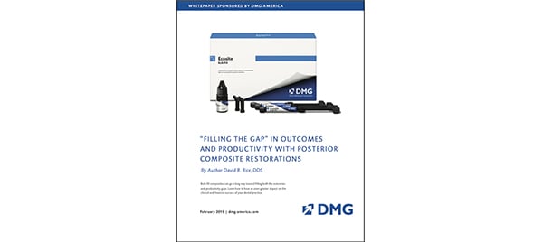
Free White Paper
Bulk fill composites can go a long way toward filling both the outcomes and productivity gaps. Learn how to have an even greater impact on the clinical and financial success of your dental practice with this whitepaper, “Filling the Gap” in Outcomes and Productivity with Posterior Composite Restorations.
Live Patient Botox Therapy and Frontline TMJ courses at ADA FDI 2019
Join us in San Francisco at ADA FDI World Dental Congress, Sept. 4-8, to learn Botox and Orofacial Pain treatment techniques that you will be able to immediately implement into your practice, led by Dr. Louis Malcmacher and his team at the American Academy of Facial Esthetics. Register now!
Advertisement

|
|
